KFJ goes DMI: We Upgraded our MV Imaging Detectors
In Dec. 2015, we replaced the original aS1000 (IDU20) MV detectors on our TrueBeams with Digital Megavolt Imagers (DMI, aS1200). From the outside, the TrueBeam looks the same. The change is hidden behind the MV imager's cover:
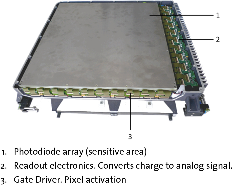
(Digital Megavolt Imager with cover removed.1)
The upgrade took less than one day per machine. The old detectors had used a digitisation unit (DU) to prepare the images for the XI. The DU is not necessary anymore, since the DMI is already "digital". A fiber optic cable now runs directly from the imager to the XI in the control room.
Clinical Advantages
At typical clinical detector distances of 160 cm, most tangential breast fields were cropped by the older IDU20 detector, which had a non-square detector size of 40 cm (width) x 30 cm height and an image size of 1040 x 768 px.
The larger area of the DMI is now square ( 43 x 43 cm for single images). This offers the possibility to image larger field lengths at the same imaging distance. Resolution is also slightly improved. Image size is now 1280 x 1280 px for single images.
Advantages for Dosimetry and QA
The detector area which can be used for dosimetry measurements (integrated images) is a little smaller than for single images: 40 x 40 cm (1190 x 1190 px). The outer frame is proably cropped to exclude scatter effects at the edge of the detector.
The DMI allows for much higher dose rates than the IDU20. Saturation would occur at about 3200 MU/min, but this is clinically not achievable. Portal Dosimetry can now be performed with 10FFF at isocenter, using the full dose rate of 2400 MU/min.
Other dosimetric advantages are better dose linearity (0.5% compared to 1.5% for IDU20), improved backscatter due to an integrated lead plate, and reduced ghosting.
As a drawback, we had to configure Portal Dose Image Prediction "the old-fahioned way" (measuring AIDA-patterns, output factors, etc.), because Varian has not released (yet) Portal Dose Pre-Configuration (PDPC) for the combination TrueBeam 2.5 + HD120 + DMI. For five photon energies, there is quite some work to do!
Clinical Example - 10FFF
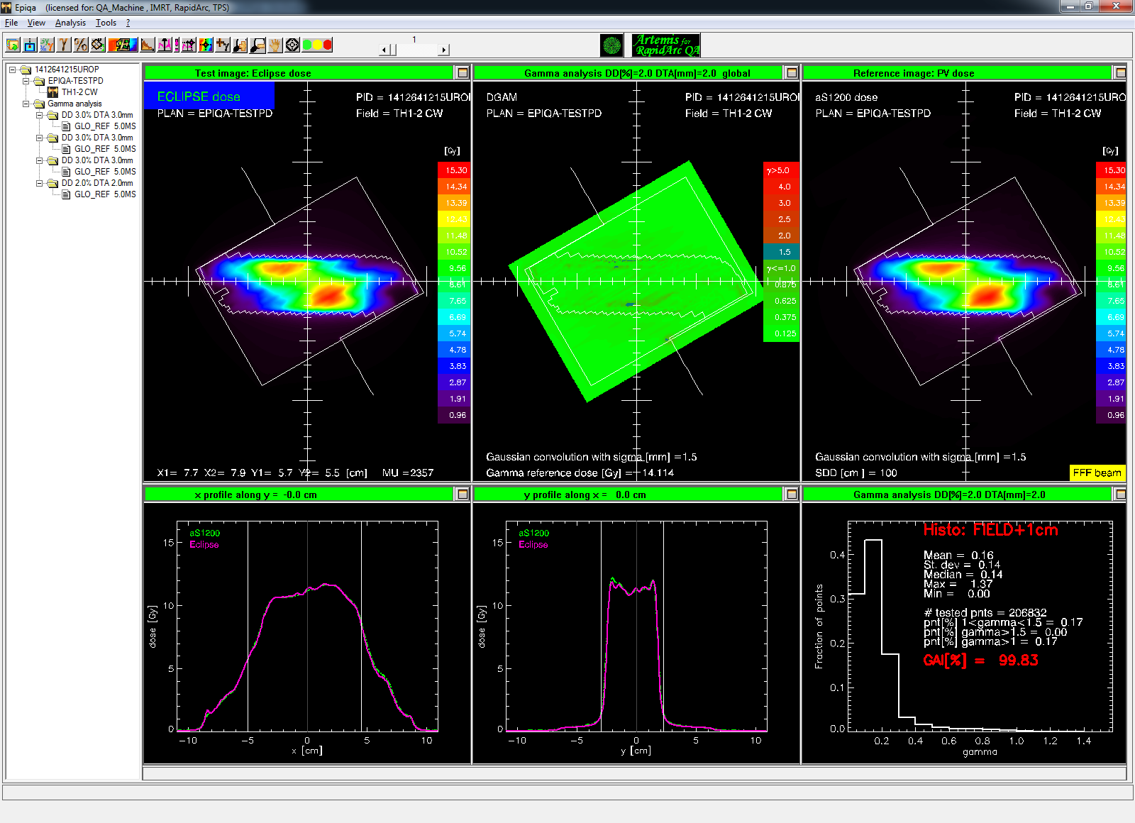
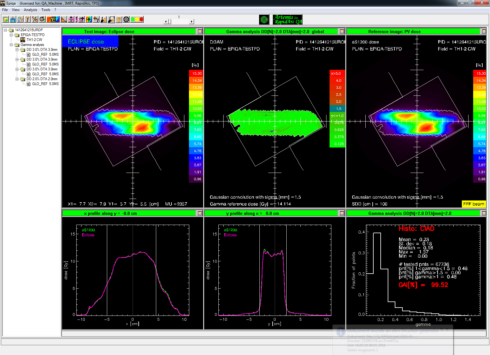
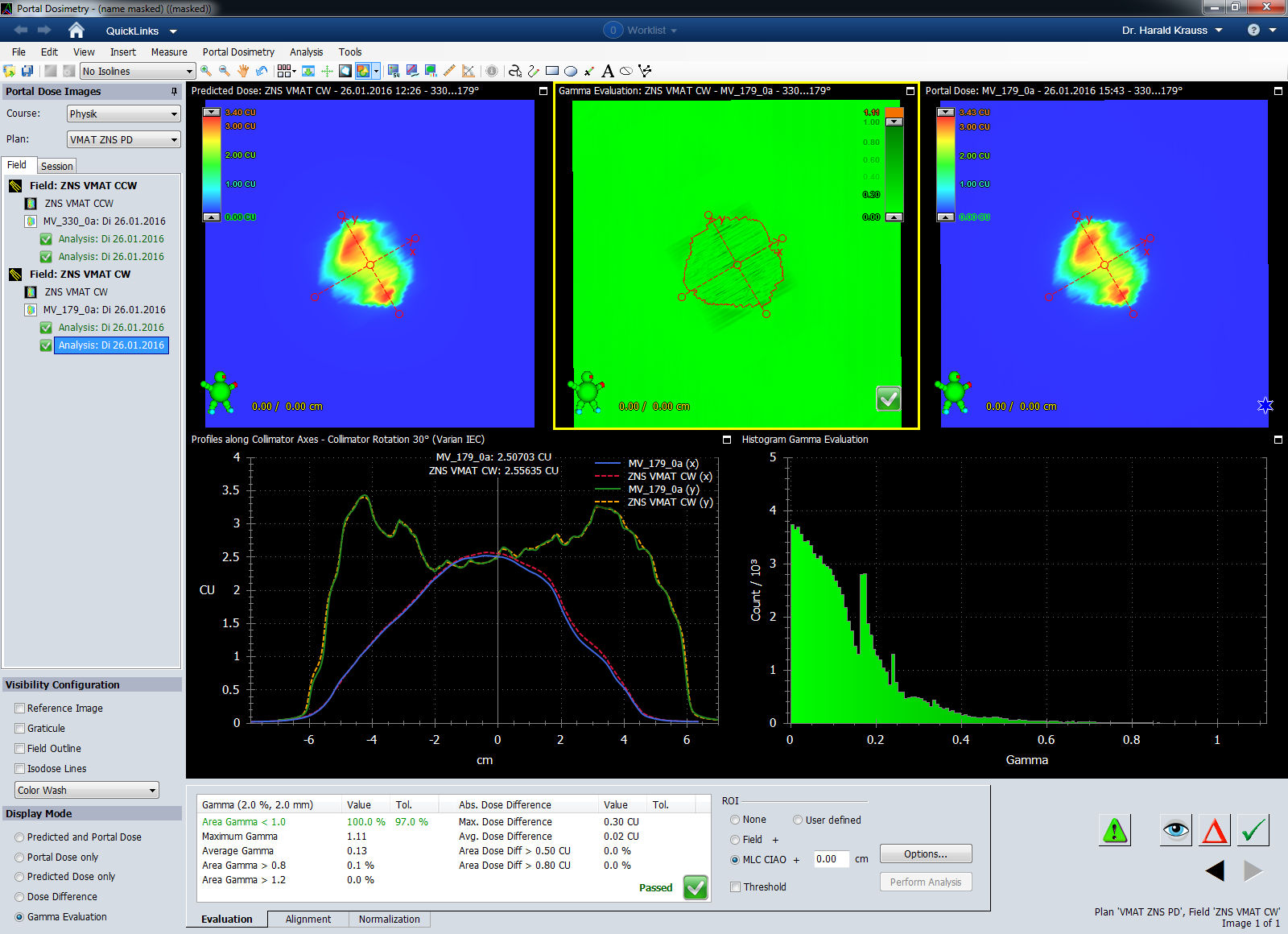
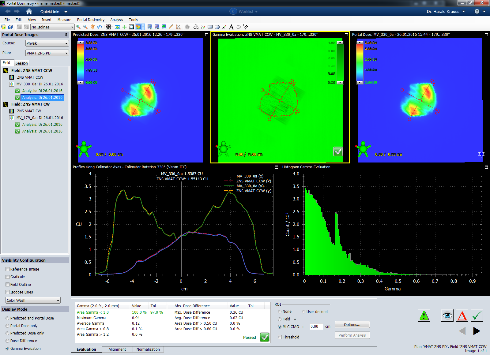
The following example shows a 1 x 8 Gy palliative treatment of Th 1-2 in a single arc, 10FFF energy, 2400 MU/min and 2357.2 MU total, planned with Eclipse 13.6 (AAA 13.6.23). This plan shall be verified with Portal Dosimetry:
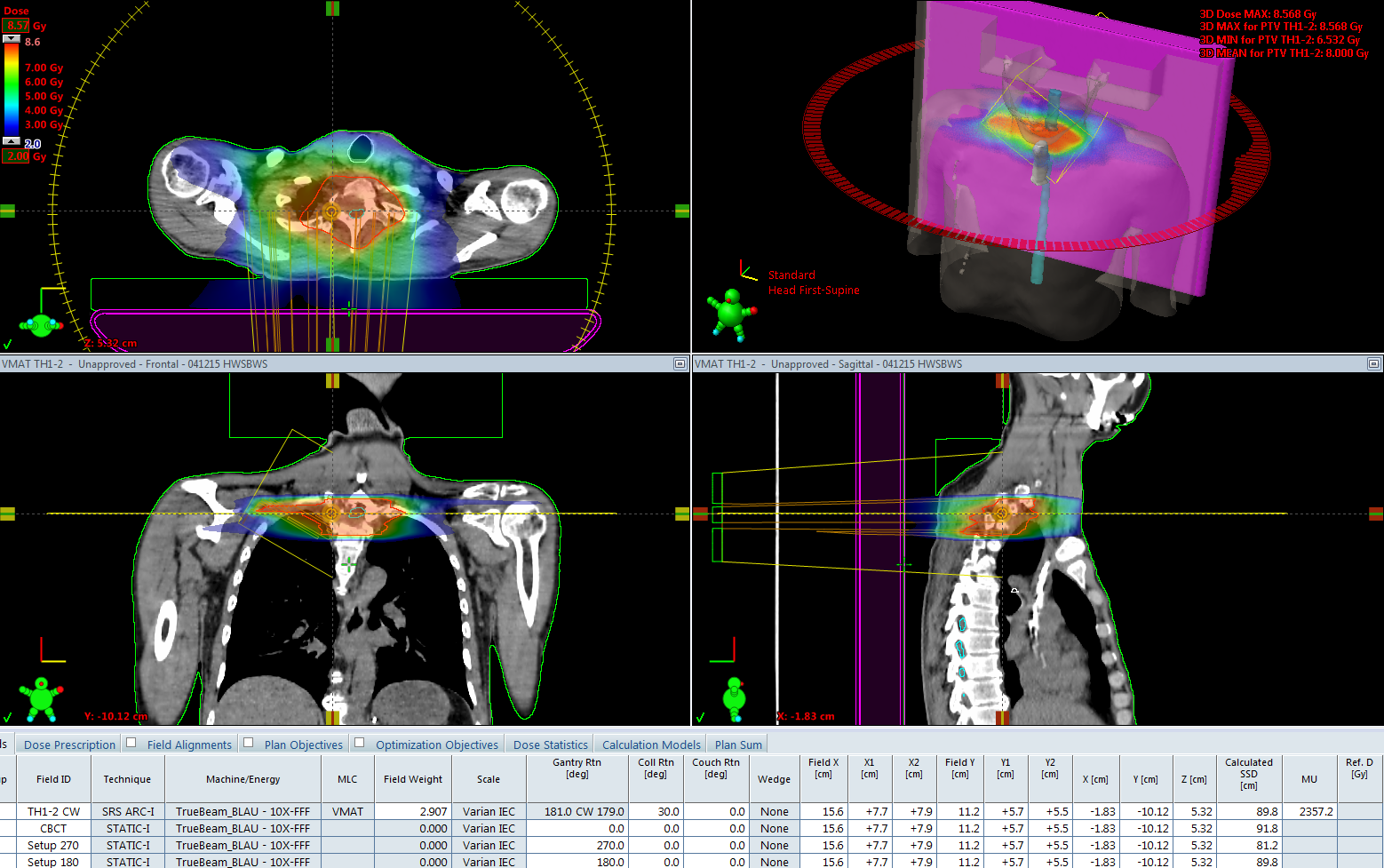
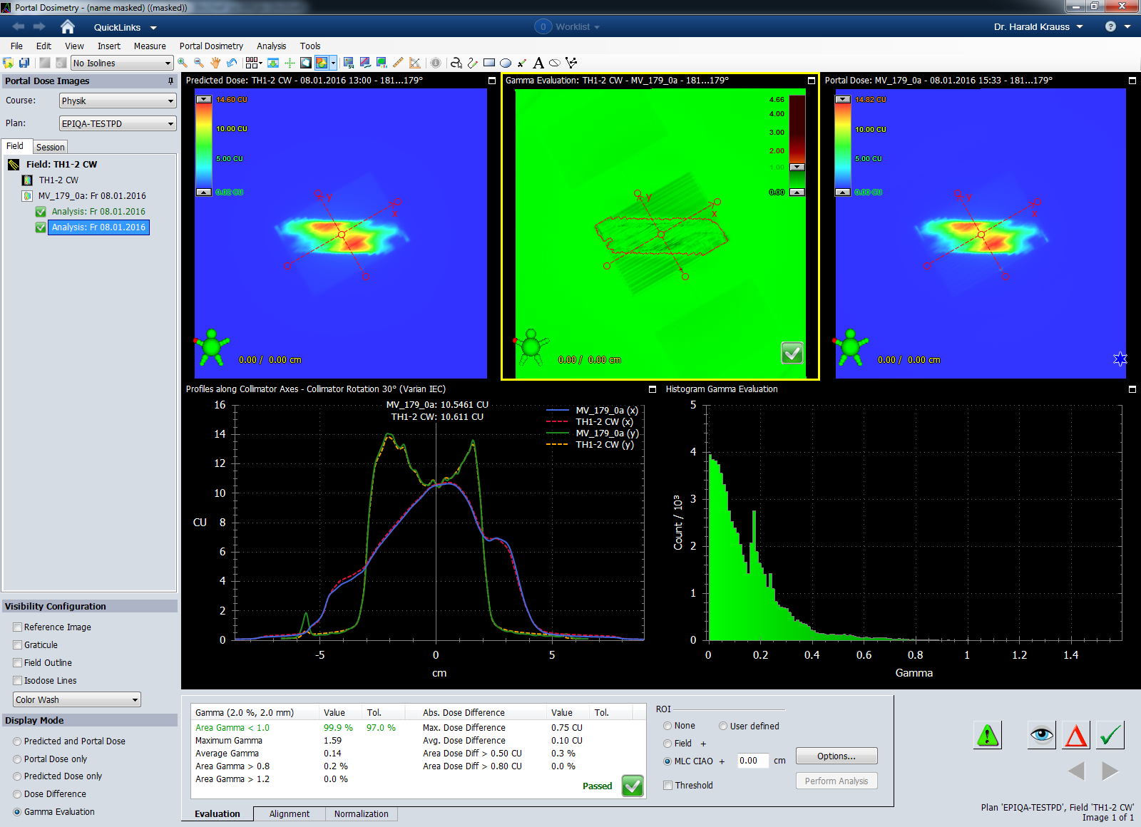
A verification plan was calculated using the Portal Dose Image Prediction (PDIP) algorithm. The arc was measured with te DMI imager at isocenter distance and analysed using the 2%/2mm Gamma criterion. In the next screenshot, the ROI is set to Field + 1cm:
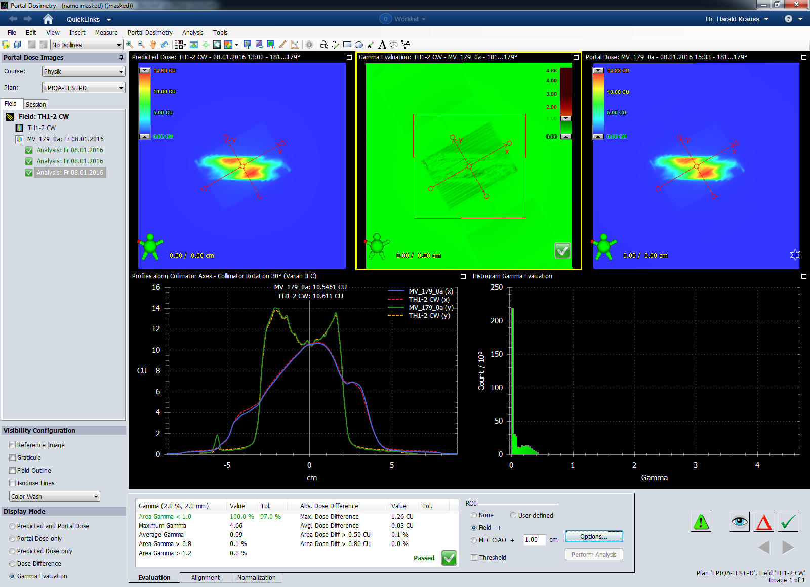
If the Complete Irradiation Area Outline (CIAO) is used as ROI, the results are similar.
The same image is then analysed with EPIQA2, first with the ROI set to Field + 1cm,
and again using the CIAO.
Note that the rotation of the Field ROI is different in the Varian Portal Dosimetry system and in EPIQA. In EPIQA, the ROI rotates with the collimator, which leads to the expected analysis area. In VPD, the ROI is a rectangle which does not rotate and which encloses the field as defined by the jaws, plus margin. A rather unnatural definition of the ROI.
Only the CIAO ROI covers the same area in both analysis systems. This is one of the reasons why we routinely only use the CIAO to document the Portal Dosimetry analysis results. Read more about it in the blog entry from May 2015.
Clinical Example - 6FFF
For the second flattening filter free energy, 6FFF, portal dosimetry results also improved dramatically. Outside the CIAO area, profiles measured with aS1000 were often to low compared to calculation (using EPIQA). This problem does not exist anymore.
Here is an example of two VMAT arcs of the brain, 6FFF, 1400 MU/min, again measured at isocenter distance. With the 2%/2mm criterion, the gamma agreement index for both arcs is 100% over the whole detector area:
Notes
1from Varian manual "TrueBeam Technical Reference Guide - Volume 2: Imaging, P1005924-001-A".
2EPIQA version 4.1.1 is needed for DMI and Truebeam 2.5.