A typical high-energy example
As we have seen on the previous page, the MIX mode is not ideal for 15MV. This is also the reason why the agreement was rather poor, despite the improvement which was achieved by using the Gaussian convolution functionality.
DMAX mode should be used for higher energies
It is better to use DMAX mode for energies higher than 6 MV. As one can see in the following example, the additional buildup plate provides scatter conditions on the sensitive detector layer which are closer to the conditions at water dmax than the scatter conditions in 8 mm depth of the "naked" EPID. In other words, the additional buildup material added at the time of measurement adds some "natural blurring" to the image. Postprocessing by Gaussian convolution becomes less important, but can still improve the agreement.
The calculation was done with the typical grid setting of 0.125 cm. This gives a 1:1 match between calculation grid points and EPIQA calculation points. AAA version was 10.0.28, EPIQA version 2.2.1. The Gaussian convolution radius of 2.0 mm was chosen. This gives visually the best match of spatial detail between the Eclipse and the EPIQA images. The manual alignment feature of EPIQA 2.2.1 was used to shift the measured matrix by 1 pixel, if necessary.
The volume treated is a very large rectum tumour. The plan has five Gantry angles, all fields are split into carriage groups.

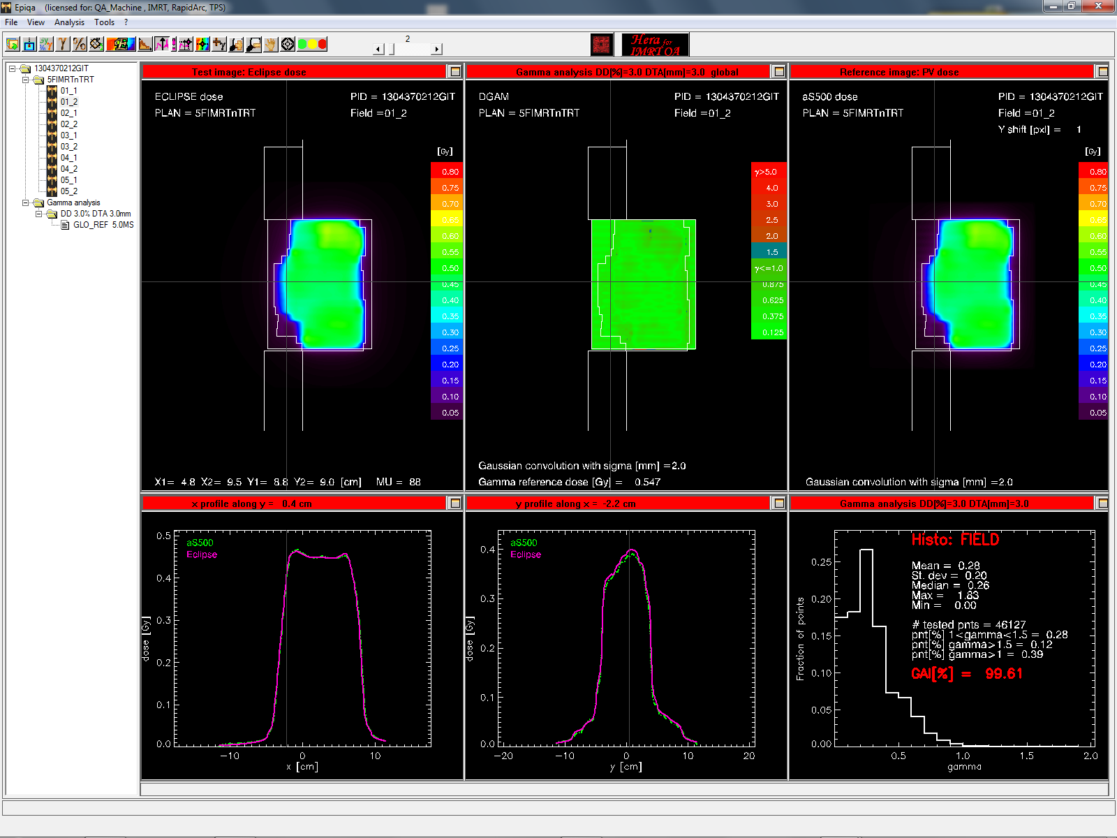


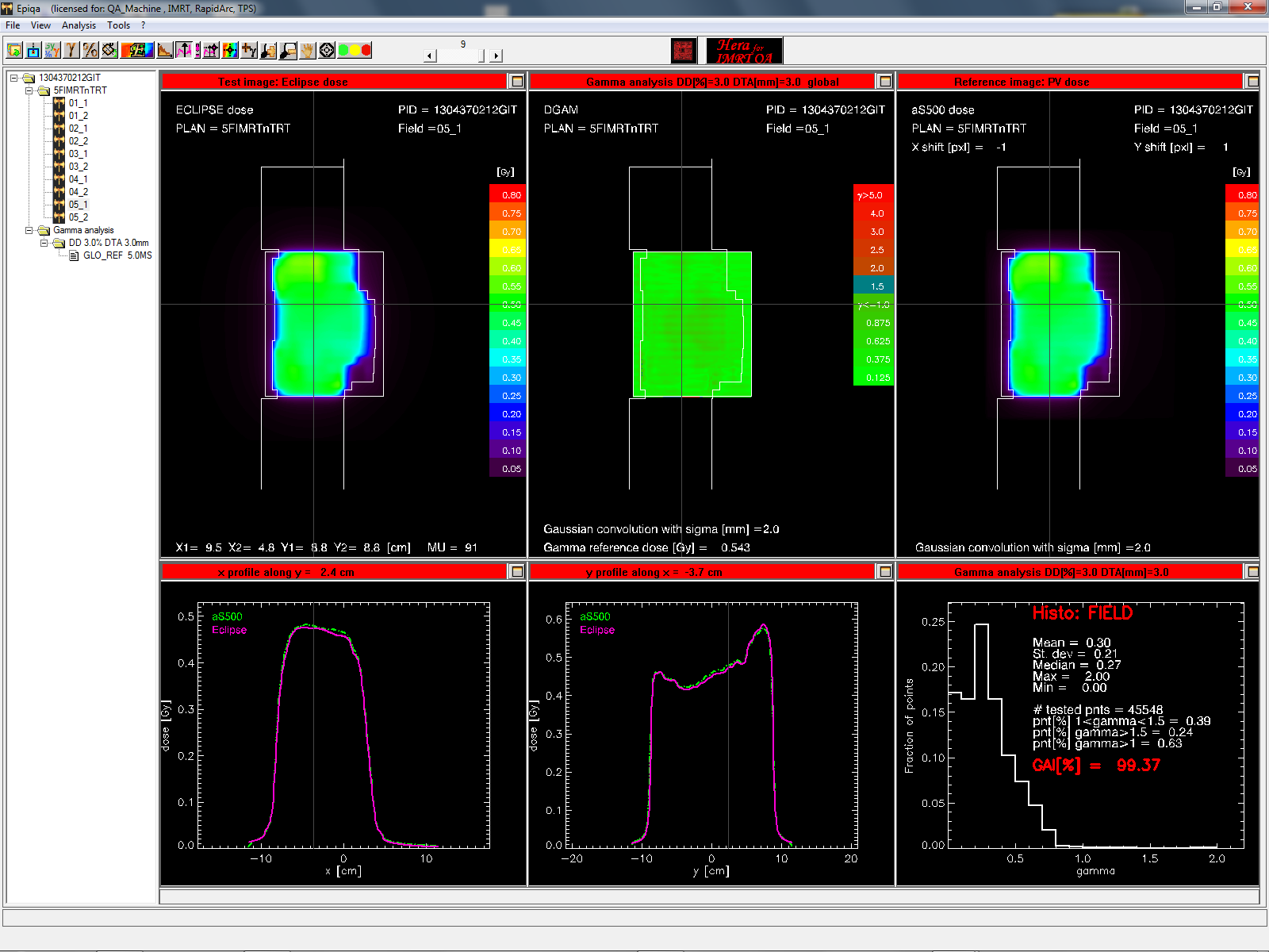
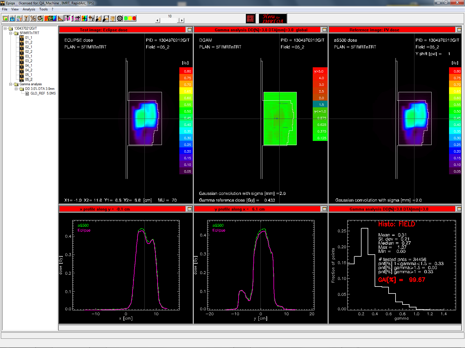
We use a 15 mm slab of PMMA for 15MV. Therefore, when in DMAX mode, dose planes in Eclipse have to be exported at 2.6 cm depth (SDD 105 cm). It is not necessary to measure in true dmax when in DMAX mode!