Results (3)
Gantry Angle Dependence
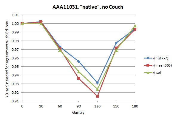
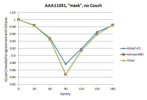
In the following graph which shows k(user) versus gantry angle for AAA and the native structure set, three different methods of finding the k(user) factors are displayed. If one evaluates a histogram over the central 7x7 chambers in VeriSoft, one gets a slightly different result than when the average dose of chamber 365 or the point dose in the chamber center is taken, especially if the calculation phantom is inhomogeneous like the native set. See the planning study in the first Results section.
(Fig.19)
Since the absolute level of k(user) is not important, we normalized to k(user)@gantry0°.
Although the angle resolution is rather coarse (30°), the message is clear: using the native scan of the OD/OP tandem for VMAT verification may be problematic. Over the half arc from 0° to 180°, k(user) for the C365 chamber changes by over 8%, and is smallest at 120°.
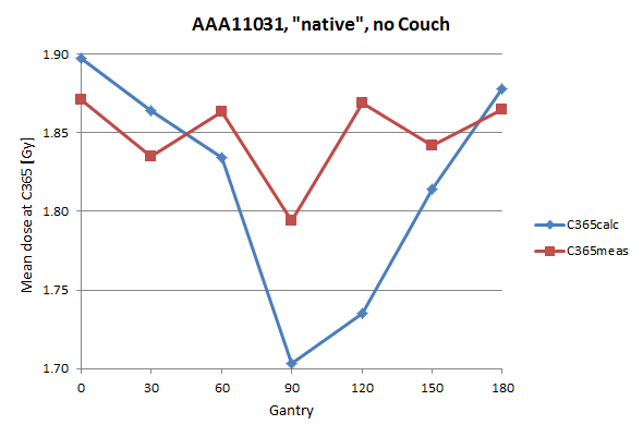
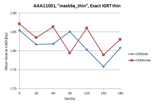
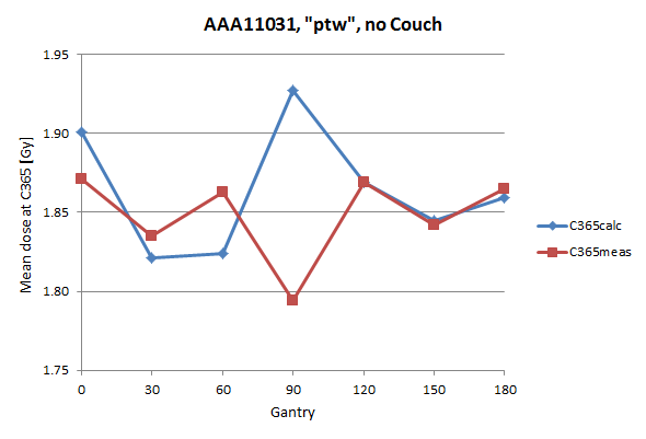
Mean calculated dose and measured dose of C365, as they contribute to k(mean365), are plotted separately in this figure.
When the dose plane through chamber center is exported, this plane is only 2.5 mm behind a strong bone-like inhomogeneity. The question is whether AAA is able to handle such build-up/build-down situations correctly. Remember the strong effect a change in calculation grid size from 1.5 to 1.0 mm had on the AAA calculated dose profiles. The more inhomogeneous a phantom is, the higher the uncertainty in dose calculation (most algorithms work well only in water).
Although the OP has a compensating air cavity, one also has to keep in mind that it was designed and optimized for the S29, not the OD.
The smaller the dependence of k(user) on gantry, the better. Ideally, the curve should be flat. If this cannot be acheived, at least some points should be above 1, some below (so that some cancelling of errors can take place). In the above curve, most points are below 1.
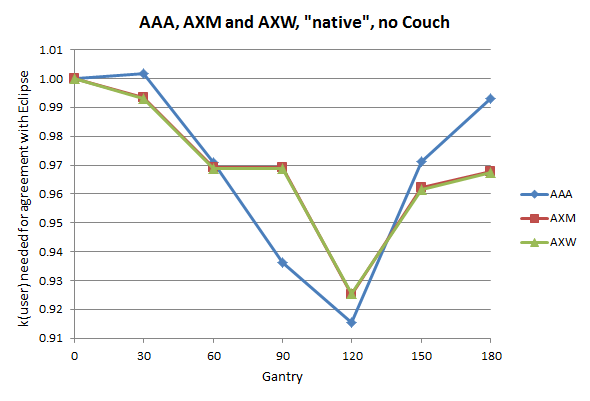
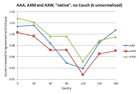
Will AcurosXB solve the problem? The next graph compares AAA and AXB. AXB has two reporting modes: dose to water (AXW) and dose to medium (AXM). The graph is for the native structure set:
(Fig.20)
The amplitude is still 8%. It looks like there is no difference between AXM and AXW. However, there is a constant shift between the two, which is removed by normalization of k(user) to 0°.
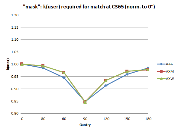
Setting the whole OD to a constant 770 HU (structure set "mask") was the first try to remove the gantry dependence, but it made things worse:
(Fig.21)
All k(user) factors are smaller than the factor at 0°, and the amplitude even increased to 15.3%. Due to the constant 770 HU over the whole OD, a beam which enters the Eclipse phantom at 90° "sees" a homogeneous dense material which is not there in reality (where chamber air volumes are sandwiched between phantom material, reducing radiological depth).
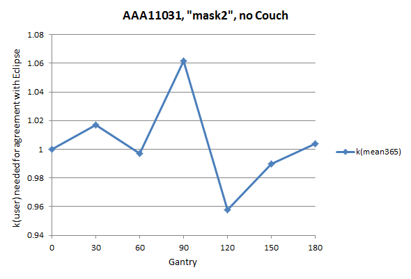
So the next try was to segment the chamber section separately (structure set "mask2"). We assigned -200HU to this structure, which is the average HU value over a VOI which only covers the 27x27 chambers, not the whole OD:
(Fig.22)
Now this looks much better. Calculated 90° dose increases, so k(user) - which is always applied to the measurement - also has to increase. On the other hand, the amplitude is still 10.4% (will it be possible to reduce this?). But at least some dose deviations have the chance to cancel out over a full arc, since some points are above 1, some below.
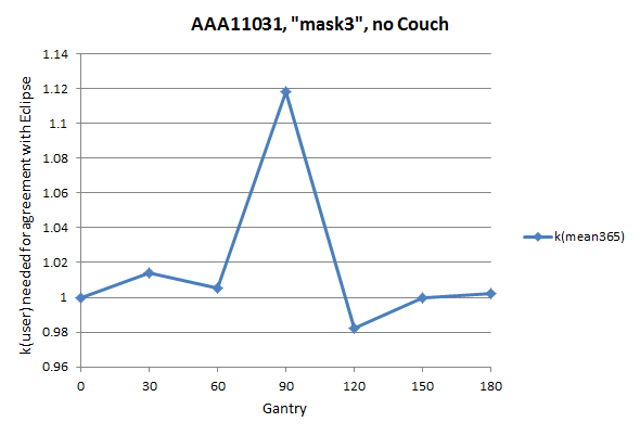
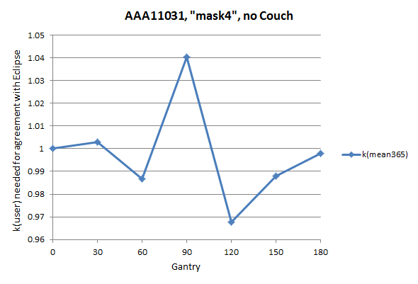
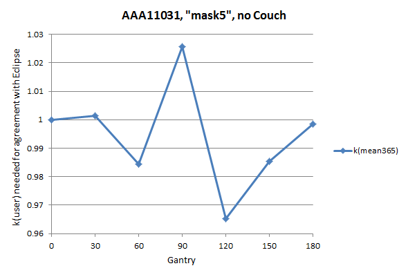
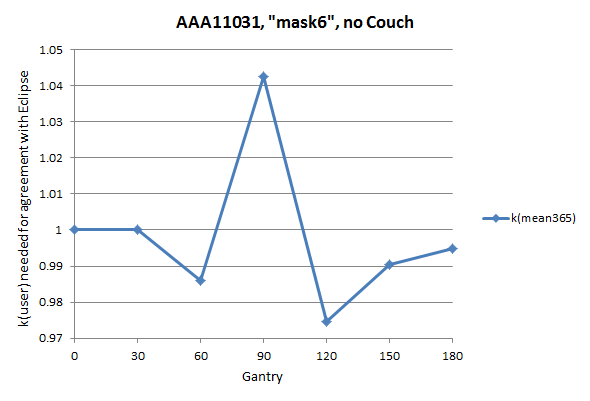
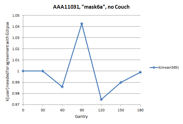
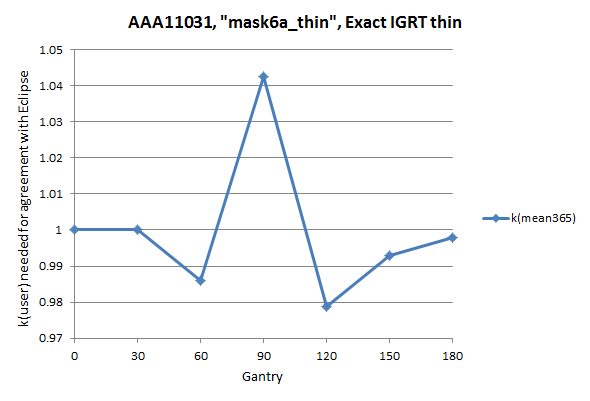
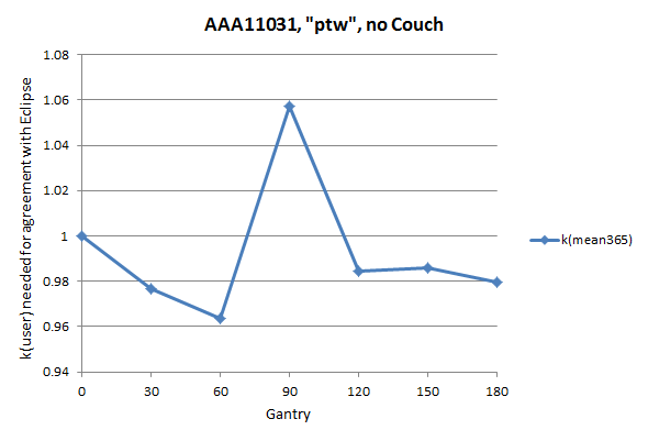
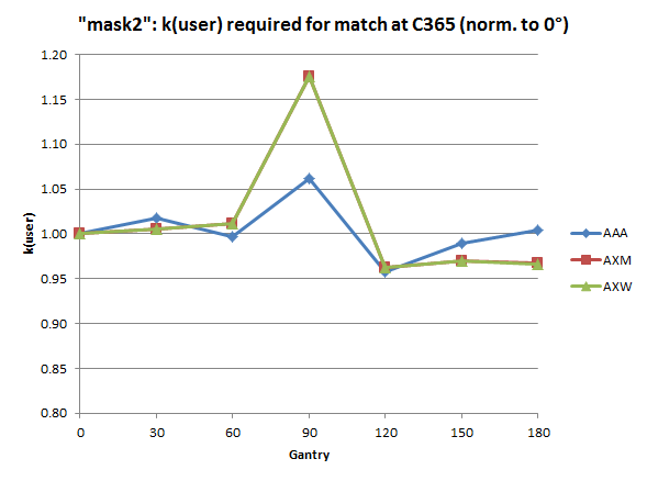
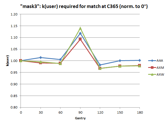
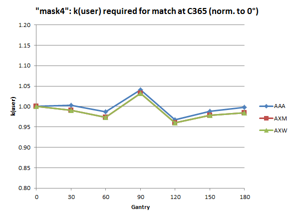
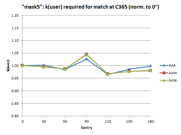
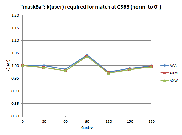
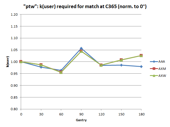
We skip some of the "manual optimization steps" which also had the goal to make the configuration more simple (if possible). This means there should not be too many structures with different densities assigned. For the interested reader, here are the k(user) vs. gantry plots for the structure sets mask3 (amplitude 13.5%), mask4 (7.3%), mask5 (6.1%), mask6 (6.8%), mask6a (6.8%), mask6a_thin (6.4%, with couch) and ptw (9.3%). All curves are for AAA. See also the separate plots of calculated and measured dose to C365 for structure sets mask6a_thin and ptw.
The AcurosXB analysis gives similar curves. Plots which compare AAA, AXM and AXW (all normalized to 0°) are here for mask, mask2, mask3, mask4, mask5, mask6a and ptw.
At this point of the optimization, structure set mask6a (mask6 with a small modification) seems to be the most promising candidate for VMAT verification. The set has the clear advantage of ultimate simplicity. It is not even necessary to scan the real phantom!
However, evaluations were only done in coarse 30° steps, and only one chamber (C365) out of a total 729 were analysed. Further insights can only be gained by measuring and evaluating real clinical RapidArc plans.