Evaluating Gantry Dependent Symmetry with EPIQA
Always looking for the most accurate yet user friendly way of doing QA, we want to present another application of the EPID which shall be used to fulfil the requirements of our national machine QA standard, ONORM S5290.
In our recent report on light field / radiation field QA, we used a phantom on the couchtop. This time, we only need the EPID itself, plus our gold standard portal dosimetry software, EPIQA from EPIdos. Current EPIQA version is 4.3.4.
EPIQA was already covered several times, mostly in the context of VMAT or IMRT patient plan QA (see for instance the patient QA results between 2013 and 2015). We use the software continually since 2011.
While EPIQA still lacks some user friendliness because the workflow involves exporting files from ARIA, its implementation of the GLAaS algorithm1 renders it physically superior to the integrated Varian Portal Dosimetry solution.
Another benefit of EPIQA is that it also works for static fields without MLC. A simple square 10x10 cm field still can't be verified with Varian Portal Dosimetry. This makes EPIQA ideal for certain machine QA tasks.
Task: Measure Symmetry at Major Gantry Angles
The Austrian standard S5290-1 requires yearly inplane and crossplane symmetry measurements at gantry angles 0°, 90°, 180°, 270° for all energies. Three points per profile are sufficient. Typically, a gantry mounted device is used for the task2. Photon field size must be at least 30 x 30 cm at isocenter.
Three points are a very crude approximation of a profile. At least for photons, there is a better solution. A Varian DMI (aS1200) image has 1190 x 1190 pixels, and we will use all of them:

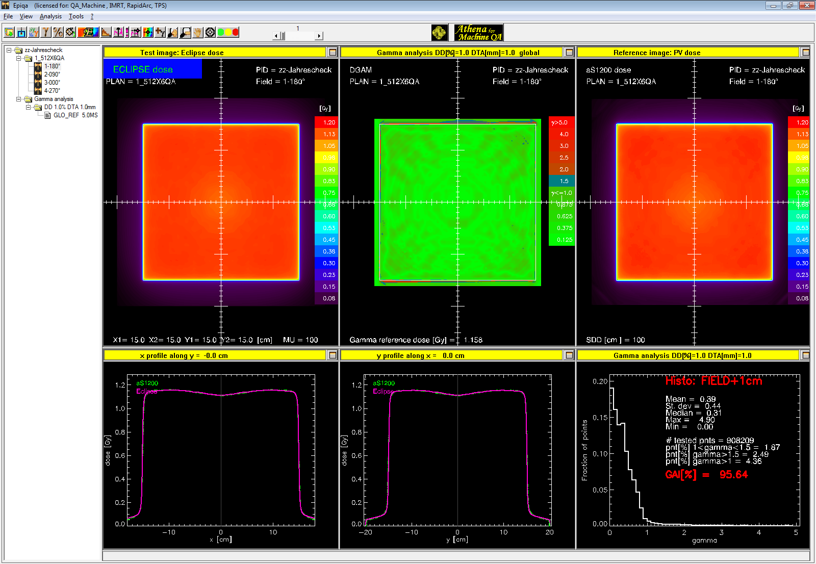
(Comparison of 10FFF Eclipse AAA plan dose and EPID measured dose. Gamma criterion is 1%/1mm global.)
The 10FFF, 30 x 30 cm field image was acquired at isocenter with full dose rate (2400 MU/min).
Our DMI upgrade in Dec 2015 in combination with the new EPIQA version make all previously published portal dosimetry results look old. And we mean: old.
Improvements in EPIQA 4.3.4 are mainly in its configuration software, which is more sophisticated than in earlier versions:

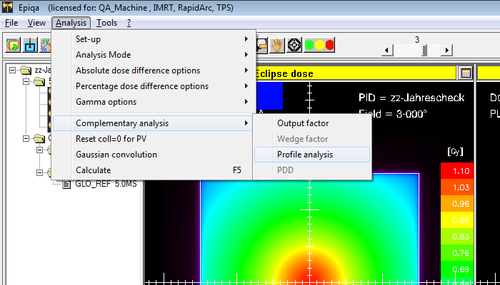
EPIQA's machine QA workflow is the same as for patient plan QA. When the software encounters fields without MLC during plan load, it switches to the Machine QA module, which is called Athena. While the above shown comparison looks definitely nice in terms of the Gamma agreement result of 98.63% for such a tight Gamma criterion, it is not what we want. We therefore select "Complementary analysis" from the menu, and then "Profile analysis":
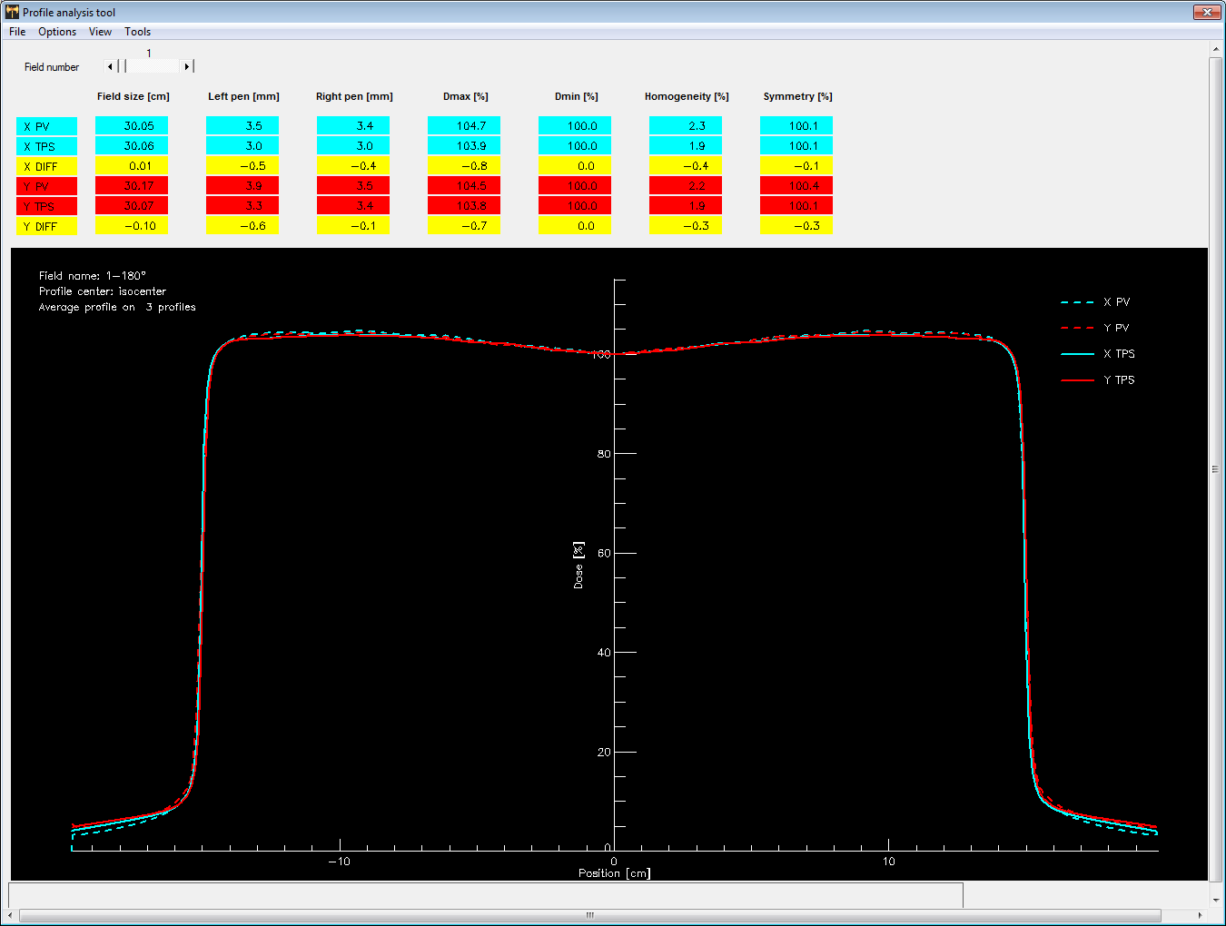
This brings up more quantitative results (click to enlarge):
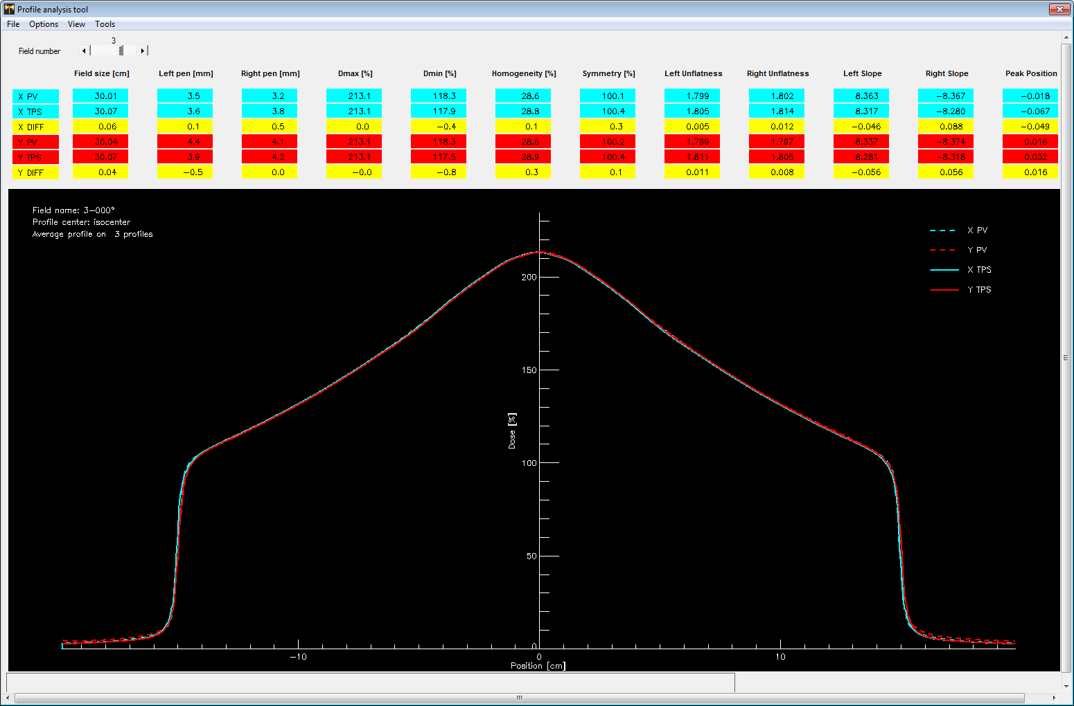
The analysis protocol can be configured, but in case of FFF, the formulas of Antonella Fogliata et al. are the only ones which make sense. A lot of data is displayed. We are mainly interested in the Symmetry results inplane and crossplane (column 7), which are 100.1% crossplane ("X PV") and 100.2% inplane ("Y PV").
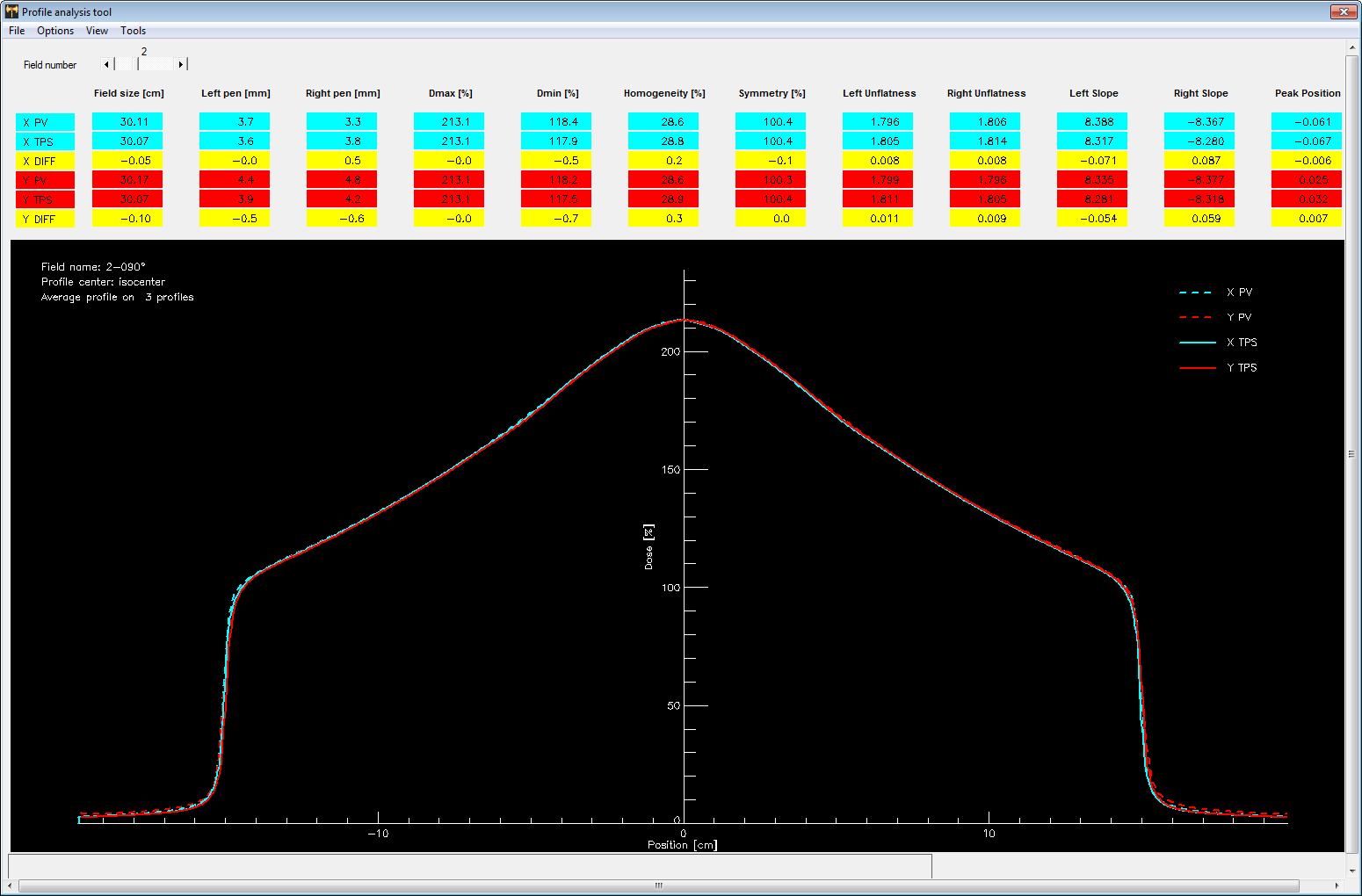
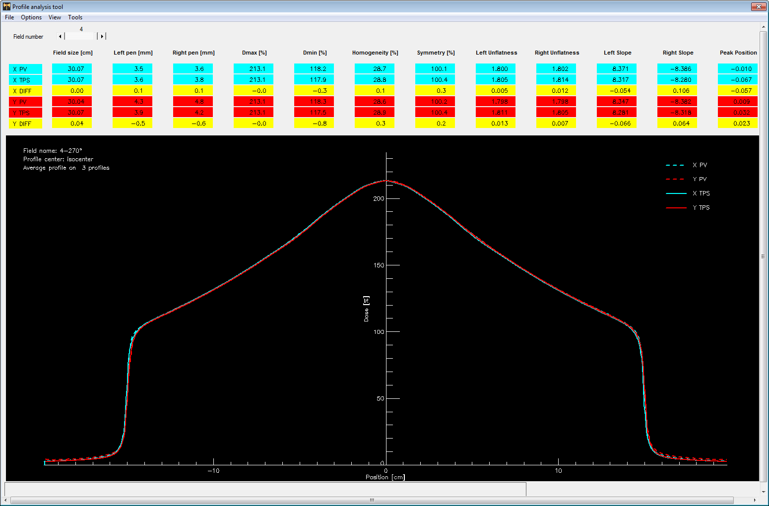
What about the other gantry angles? The results for 90°, 180° and 270° are very similar, due to the high accuracy of the TrueBeam system. The highest symmetry values we found were 100.4%, for both TPS and the EPID (PV).
After selecting some options,
the final results are presented as HTML report, which is automatically saved and can be printed:
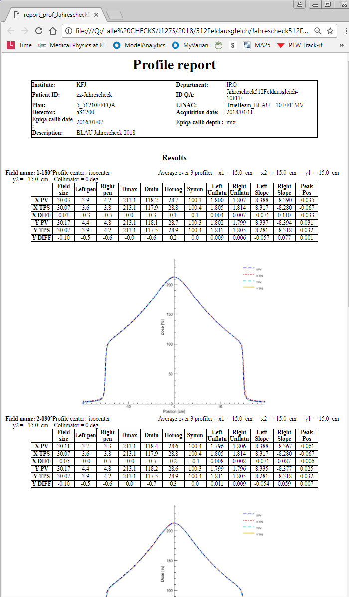
(First two profiles of HTML report. Including the plots, the report has three pages.)
For the other photon energies, the results are similar. For example, here is the 6 MV image acquired at 180° and evaluated in the same way as above (1%/1mm global):
The only pixels which fail are in the penumbra region. Keep in mind that Varian considers the jaws as fit for backup shielding but not for high precision field shaping. The Y jaws have a tolerance of 2 mm (per jaw!) at isocenter, even on the TrueBeam. This might give an explanation for the effect, which is not relevant for the task at hand.
Finally, this is the corresponding profile analysis for 6 MV, 180°:
Discussion
Ten minutes are needed on the TrueBeam to acquire the 20 + 5 images (four images per photon energy at the four gantry angles plus one 10x10 reference image for EPIQA). Another 10 minutes are required to export all images from ARIA, due to the extremely lame ImportExport application of ARIA 15.1.
On the positive side, roughly 1.5 million pixels are giving full dose information (in Gy!) on what the beam does at various gantry angles, no only 5 points. This renders EPIQA superior over all known alternatives for the described task, at least on the TrueBeam.
Notes
1 GLAaS uses empirically measured data (= integrated EPID images acquired on the user machine) and derives calibration data for the EPID pixels to convert pixel value to dose (in Gy).
2 For electrons, we still use such a device.