Portal Dosimetry is a system for the verification of IMRT plans. It has the following components:
- The portal dose image prediction (PDIP) software. The terms Portal Dose Prediction (PDP) and Portal Dose Image Calculation (PDIC) are also used. This module is part of the Eclipse treatment planning system.
- The Portal Imager (aS1000, aS500 amorphous silicon), to measure the image.
- The ARIA Portal Dosimetry Review workspace (see screenshot below), to evaluate the agreement between predicted and measured images.
If you are new to the subject, you should first read about the principle.
Clinical examples can be found here.
There are also some minor dosimetric problems.
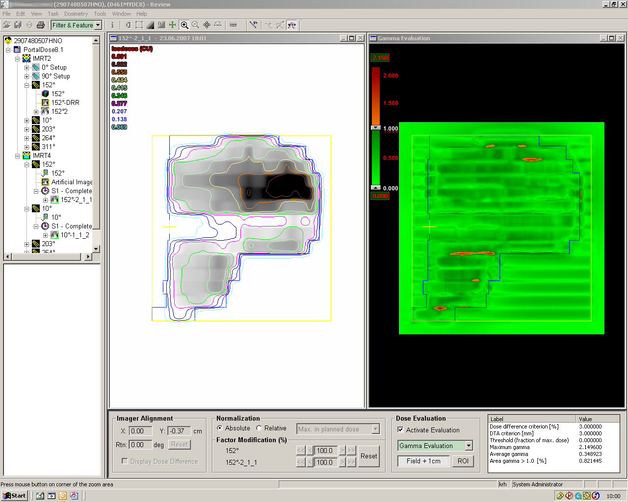
Example of evaluation in absolute units.
Left: measured image and calculated isodoses. Right: Gamma map. Pixels failing the Gamma criterion are drawn in red.
We often get asked: what is it that you measure with Portal Dosimetry? Is it dose? Or rather fluence? Hard to say. The processes inside the amorphous silicon imager are rather complicated and not comparable to ionization chamber dosimetry. To avoid the protests of hard-core dosimetry experts, the terms "dose" and the corresponding unit "Gy" is avoided by Varian in the context of Portal Dosimetry. Instead, the software manages Calibrated Units (CU). Among friends, 1 CU corresponds to 1 Gy. Why? See the page on imager calibration. For simplicity, I will therefore use the term Dose, although it is physically not correct.
We do not know what the quantity is that we are measuring, but at least we do it in absolute units!
The Portal Dose Image Prediction (PDIP) is an algorithm which is different from the algorithms used for volume dose calculation (Pencil Beam, AAA, etc.). Since the depth in the portal image detector is always the same, a 2D algorithm is used. This algorithm first has to be configured in Beam Configuration before it can be used in Eclipse. Among other things, an image of a test pattern has to be measured with the imager, and output factor tables of the imager have to be measured. This corresponds to the beam data measurements (water-phantom) needed for configuration of 3D dose-calculation algorithms. It is still under discussion whether all data have to be measured separately for every imager that is used with PDIP. The best is to check and compare.
Configuration Steps for Portal Dosimetry
- Imager Calibration
- Calibrate IMRT acquisition modes on the port imager (basic image quality)
- Calibrate dosimetry of port imager
- Prepare the Kernels
- Convert fluence test pattern to DMLC sequence in Eclipse
- Measure two images on the linac
- Measure output factors for different field sizes with port imager
- PDIP Beam Configuration
- Import all data, calculate beam data