Application of the 2D-ARRAY for Relative Electron Dosimetry
The following example shows how the 2D-ARRAY can be used for answering general dosimetry questions in a simple and elegant way.
A patient should receive
treatment with 6 MeV Electrons. The field was located close to the eye, which
should be shielded. One possibility was to use a 1mm thin slab of lead, which
is flexible and can be cut and formed easily to fit the shape of the face. The
alternative was a 1.5 cm slab of SuperStuff. We decided that measuring the absorption
directly would probably be the best solution.
The 20 cm x 20 cm Electron applicator and the 2D-ARRAY were set up on the linac. No
buildup was used, therefore the measuring depth was 5mm for the open field (inherent
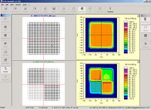
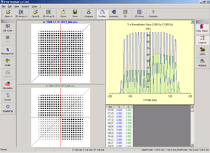
buildup of the array). With VeriSoft, I first made an open field measurement
(A) with 200 MU and 6 MeV, then a measurement with the upper right quadrant of
the field covered with SuperStuff and the lower left quadrant with the slab
of lead (B). In this way, both materials can be measured simultaneously.
Click on images to enlarge.
 |
 |
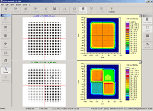
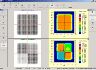
When the cursor is moved over the SuperStuff- and lead-quadrants, the local percentage dose difference is displayed in the bottom row.
 |
 |
Results: The attenuation under SuperStuff and lead was about 36% and 62%, respectively. In the open field quadrants, hot spots can be seen (red color), which are caused by scatter near the edges of the absorbing material. The increase is +1.7% near the lead edge, and +6% near the SuperStuff edge.
Performing the same measurements with a ionization chamber would give less information (single point!) and take more time.